Dental radiology is a branch of dentistry that supports diagnosis, treatment planning, and follow-up processes through the imaging of the tooth, jaw, and surrounding tissues. The technologies used in this field enable a detailed examination of tooth morphology and bone tissues. Radiological imaging plays a critical role not only in the detection of dental caries but also in implant planning, assessment of temporomandibular joint disorders, impacted teeth, and cysts and tumors.
Scope and Importance of Dental Radiology
Radiological examination is an essential diagnostic tool that complements clinical examination in dentistry. Problems in areas that cannot be seen by the naked eye are detected through radiographic images.
As a result:
- Accurate diagnosis and treatment planning are achieved.
- Unnecessary interventions are prevented.
- Operational risks are reduced.
The fundamental goal of dental radiology is to provide maximum information with minimal radiation. Today, digital systems have reduced radiation doses by up to 70% compared to conventional radiography.
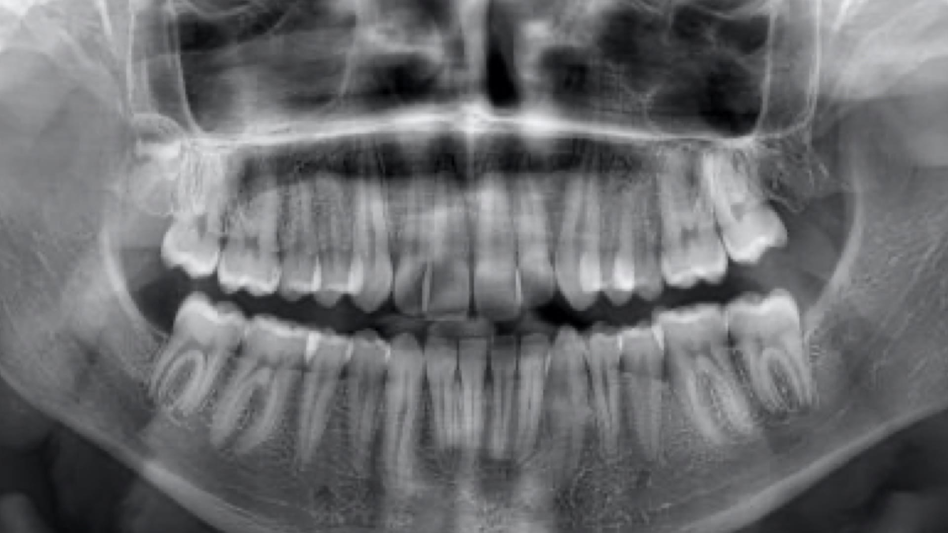
Panoramic X-Ray (OPG)
A panoramic X-ray provides the ability to evaluate the entire maxillofacial region in a single image. In this system, teeth, jawbones, sinuses, joint areas, and surrounding tissues are examined as a whole.
Areas of Use:
- Determination of the position of impacted teeth
- Detection of jaw fractures and cysts/tumors
- Evaluation of periodontal bone loss
- General analysis prior to implant planning
- Orthodontic treatment planning
Advantages:
- Displays the entire oral anatomy in a single image.
- Fast, painless, and involves low radiation exposure.
- Ideal for general evaluation before treatment.
Panoramic X-rays are generally taken every 6–12 months for a check-up or as a routine control before treatment.
Cone Beam Computed Tomography (CBCT)
CBCT is a three-dimensional imaging technology used in dentistry. It provides much more detailed information than conventional panoramic X-rays and is widely used in implantology, endodontics, oral and maxillofacial surgery, and orthodontics.
Advantages of CBCT:
- Three-dimensional measurement and volumetric analysis
- Evaluation of the nerve canal, bone thickness, and density
- Precise implant planning
- Analysis of the association between impacted teeth and surrounding tissues
- Determination of the size and location of cysts and tumors
CBCT is particularly used to measure the bone height and thickness of the area where the implant will be placed with millimetric accuracy. This helps minimize risks before the surgical procedure.
Technical Features:
- Creates a 3D volume using a rotating cone-beam X-ray source.
- Provides high image resolution with a low radiation dose.
- The obtained data are digitally stored and used in planning software.
Other Dental Imaging Techniques
In dental radiology, there are several additional imaging methods besides panoramic X-rays and CBCT:
- Periapical X-ray: Shows the details of a single tooth or a small area. It is used in root canal treatment and examination of root morphology.
- Bitewing Radiograph: Effective in evaluating caries on adjacent tooth surfaces and the fit of fillings.
- Cephalometric X-ray: It is used particularly in orthodontics to analyze jaw position and skeletal relationships.
- Digital intraoral scanning: It does not involve radiation and is used for the digital measurement of surface structures.
These methods can be used either alone or in combination, depending on the patient's needs.
Radiological Safety and Radiation Dose
Radiation exposure is significantly reduced through the use of digital sensors in modern dental radiology systems. The effective radiation dose from a panoramic radiograph is minimal and represents only a small fraction of the daily natural background radiation.
Safety Measures:
- Use of a lead shield and thyroid shield
- Short exposure time with digital systems
- Avoidance of unnecessary repeat exposures
- Special protection protocols for children and pregnant patients
In terms of radiation exposure, dentistry is considered one of the lowest-risk fields in medicine.
Dental radiology is an indispensable diagnostic and treatment planning tool in modern dentistry. While panoramic radiography provides information about the overall structures, CBCT technology enables three-dimensional analysis with millimetric accuracy. These methods offer significant advantages for dentists in establishing accurate diagnoses, safe surgical planning, and effective treatment processes. Thanks to low-dose digital systems, a safe and efficient imaging process can be achieved for patients.